Cornea
The cornea is a transparent and thin layer like a contact lens located upfront of the eyeballs. The most important and have to be preserved quality is the transparency of the cornea.
A potential donor willing to help others after they pass away will only give one thin cornea layer, NOT the whole eyeball.
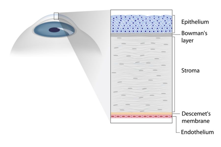
The cornea consists of a few layers, such as:
1. Epithelium: The most superficial layer protecting the cornea from foreign body contamination. A person with epithelium injury will experience pain and watery eyes. This layer is easily infected if there's a wound or scratch caused by fingernails unintentionally. This is the utmost cornea layer functioning as a barrier
2. Bowman Layer: Layer in which epithelium adheres. One will have symptoms of recurrent scars (RCE) – recurrent corneal erosion if there's a disorder in this layer
3. Stroma: The thickest layer encompassing 85% of the cornea, composed of collagen fibres. Viral stroma infection causes one to be sensitive or dazzled by the lights (photophobia)
4. Descemet membrane: A tissue in between stroma and endothelium
5. Endothelium: The deepest layer with the critical role of maintaining clarity or cornea transparency, consists of thousands of endothelium cells; the number has to be observed timely when one is diagnosed by an ophthalmologist suffer a genetic cornea disorder called corneal dystrophy.

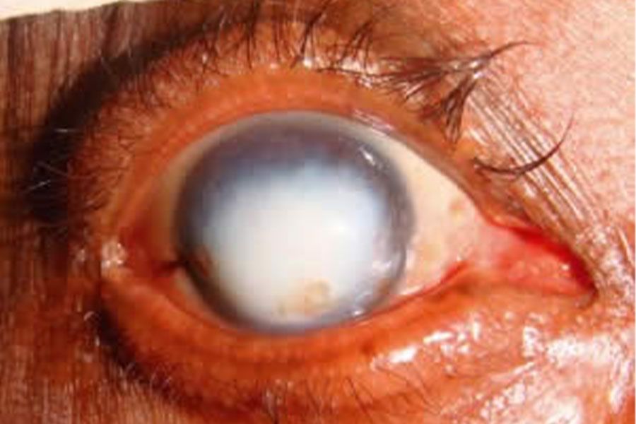
Cornea Blindness
One's eyesight can be impaired or worse if a disorder of cornea transparency is present. This disorder is caused by infection, hereditary, infection scars or wounds, and chemical, trauma and mechanic trauma.
Emergency Cornea Disorder
is a condition in which the cornea experienced disorder, so infection initially located in the cornea can spread to the whole eyeball (a condition in impending perforated ulcus), and the presence of torn cornea causes the disease to easily invade if a closure is not immediately done
Common Cornea Disorders Requiring Transplantation
Cornea Infection (Keratitis): Someone will complains photophobia during keratitis.
Some possible etiology of this condition include:
1. Virus: A well-defined typical presentation depending on the viral aetiology
2. Bacteria: Infected lesion has a blurry border compared to a virus, familiar in contact lens users with bad hygiene status, sometimes the infection gets severe, causing pus inside the eye
3. Fungi: Infection in people who wash their eyes using betel water, breastmilk, or over-the-counter steroid eye drop to cure red eye. The infected lesion will have a blurry border with distinct characteristic
4. Amoeba/germs: caused by the usage of contact lenses, sometimes used during swimming.
Cornea Dystrophy is an acquired cornea disorder in one family or several family members with a similar condition. The unique character is a distinct presentation with a specific pattern (depending on the impacted cornea layer). Someone with dystrophy disorder will experience blurry vision and photophobia/ light sensitivity. A few common dystrophies include:
o Anterior Basement Membrane Dystrophy (ABMD): one of the reasons people complain of recurrent wound/erosion (RCE)
o Granular/lattice dystrophy: stroma disorder with a unique presentation
o Keratoconus: Cornea thinning should be suspected, especially if one has astigmatism/high eyesight cylinder symptoms that can't be corrected maximally using spectacles. One can argue to have visited the eye centre several times, but none can prescribe the right size spectacles
o Bullous Keratopathy: One complaining about frequent pain dan blurry vision after cataract operation
o Fuchs Dystrophy: Insufficient endothelium cells to keep the cornea clear
Source :https://www.cehjournal.org/article/corneal-blindness-prevention-treatment-and-rehabilitation/
.jpg)
Cornea transplantation can be a procedure in which the damaged cornea will be replaced with an excellent healthy cornea. After successful transplantation, the patient is expected to have regular sight, similar to pre-injury.
The corneal transplantation technique is divided into 2 category:
- All cornea tissue transplantation (Penetrating keratoplasty)
- Half cornea tissue transplantation (Lamellar keratoplasty)
Type of corneal transplantation :
- Penetrating Keratoplasty
Transplantation operation in which the new cornea replaces all cornea layers

- Lamellar keratoplasty
Transplantation operation in which only half of the damaged cornea will be replaced. Lamellar keratoplasty is furtherly divided into three kinds such as::
1. Anterior Lamellar Keratiplasty (ALK):
If the disorder only happens in the front layer of the cornea. The unaffected part is maintained

2. Browman Layer Transplantation (BLT):
Only the upfront thin layer is replaced. Both ALK and BLT are performed if dystrophy abnormalities like keratoconus are present. The difference is there are no stitches made in BLT, while there are few in ALK
3. Endothelial Keratoplasty/EK, is divided into two kinds such as:
- Descemet's Striping Automated Endothelial keratoplasty (DSAEK): Back layers of cornea replacement/endothelium from stroma till endothelium
- Descemet Membrane Endothelial Keratoplasty (DMEK): Back layer cornea replacement in which it only replaces endothelium and Descemet membrane without stroma
Lamellar EK procedure will be done in people with cornea abnormalities on the back layers. The front layer of the cornea is undisrupted; therefore, no procedure is needed. This is usually done to people with Fuchs disorder or bullous keratopathy. Both are complications from a cataract operation – the sight is still blurry and sometimes painful after the procedure

The operation technique is decided by an ophthalmologist based on the patient's cornea condition. Lamellar keratoplasty offers some benefits, like lower donor rejection risk and shorter recovery time than whole cornea replacement or penetrating keratoplasty.