

 INA

What Is Microphthalmia? Learn About Its Causes and Symptoms!
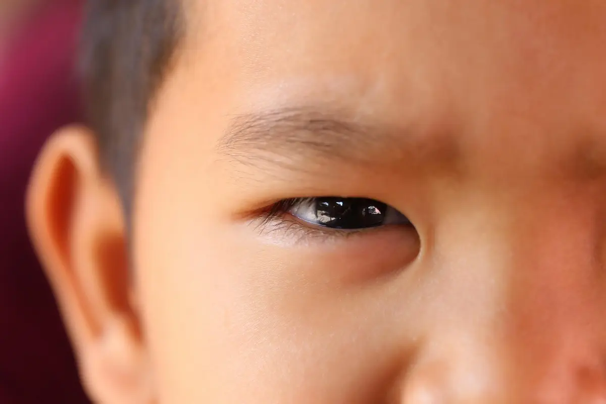
The size of a baby’s eyes generally develops normally while still in the womb. However, in some cases, a baby may be born with one or both eyes smaller than they should be.
This condition, known as microphthalmia, is a congenital disorder that can affect vision and often requires early evaluation and treatment by an ophthalmologist. To learn more about microphthalmia, read the following explanation.
What Is Microphthalmia?
Microphthalmia is a congenital condition in which a baby’s eyes do not develop fully, resulting in eyes that are smaller than normal. This condition is also known as small eye syndrome.
It can affect one eye (unilateral microphthalmia) or both eyes (bilateral microphthalmia). If it affects only one eye, the affected eye appears smaller than the healthy eye. Meanwhile, if it affects both eyes, both appear smaller than normal.
The Differences Between Microphthalmia and Anophthalmia
Microphthalmia and anophthalmia are two congenital abnormalities that affect the development of the fetus’s eyes. The main difference lies in the degree of eye formation and the size of the eyeball.
Microphthalmia is a condition in which one or both eyeballs form, but their size is abnormal (too small). On the other hand, anophthalmia is a condition in which one or both eyeballs do not form at all, so that the eye sockets appear empty.
The following is a comparison table:
Causes of Microphthalmia
To date, the exact cause of microphthalmia remains unknown. However, this condition is believed to result from a combination of genetic and environmental factors that affect fetal eye growth during pregnancy.
Some known causes and risk factors include:
- Genetic abnormalities, namely changes (mutations or variants) in genes involved in eye formation during fetal development. Currently, more than 90 genes are believed to be associated with microphthalmia. Among these, mutations in the SOX2 and OTX2 genes are the most commonly identified causes, particularly in cases that affect both eyes and present with severe symptoms.
- Chromosomal abnormalities, which are changes in the number or structure of chromosomes, can disrupt the development of the eyes and other organs.
- Infections during pregnancy, such as rubella or toxoplasmosis, which can interfere with eye growth in the fetus.
- Exposure to certain medications during pregnancy, such as isotretinoin (a medication for severe acne) and thalidomide, which are known to increase the risk of birth defects, including microphthalmia.
- Exposure to harmful substances or radiation during pregnancy, such as pesticides, certain chemicals, or X-rays, in amounts that can affect fetal development.
- Vitamin A deficiency in pregnant women, as vitamin A plays a crucial role in the formation and growth of the fetus’s eyes.
What Are the Characteristics of Microphthalmia?
The main characteristic of microphthalmia is that one or both eyes appear smaller than normal at birth. In addition to small eye size, some patients also typically have other abnormalities in one or both eyes (complex microphthalmia), including:
- Vision impairment: A child’s level of vision can vary greatly, ranging from poor or blurry vision to total vision loss (blindness). This depends mainly on how well the internal eye structures have developed.
- Congenital cataracts: The cloudiness formation in the eye’s lens, which causes vision to become increasingly blurry.
- Coloboma: The absence of eye tissue or tissue that has not formed properly. This condition is most commonly seen in the iris (the colored part of the eye), causing the pupil to appear slit-like and not perfectly round (like a keyhole).
- Microcornea: A condition in which the cornea is very small.
- Retinal detachment: A serious condition in which the retinal layer at the back of the eye detaches from its supporting tissue, which can lead to blindness.
- Ptosis or pseudoptosis: A condition in which the eyelid appears droopy or hangs down (drooping eyelid). In cases of microphthalmia, this drooping eyelid is often pseudoptosis. This means the condition is not caused by a nerve or muscle disorder of the eyelid, but rather by the eyeball being too small to optimally support the eyelid above it.
Since microphthalmia sometimes develops alongside certain genetic syndromes (such as CHARGE syndrome or Lenz microphthalmia syndrome), its symptoms are sometimes accompanied by physical abnormalities outside the eyes, such as a cleft lip, deformities of the hands or feet, and intellectual developmental delays.
How to Diagnose Microphthalmia
Microphthalmia can be diagnosed during pregnancy or after the baby is born. The doctor makes a diagnosis based on a medical history (anamnesis), a physical examination, and, if necessary, additional tests to determine the size of the eyeball and identify any other possible associated abnormalities.
The diagnostic process generally includes:
- Medical history (anamnesis): The doctor asks about the history of pregnancy and childbirth, including whether the mother experienced any infections during pregnancy, took certain medications, was exposed to hazardous substances or radiation, and has a history of nutritional deficiencies. The doctor also inquires about a family history of eye abnormalities or genetic disorders, as some cases of microphthalmia are linked to hereditary factors.
- Prenatal ultrasound (Ultrasonography), which can be performed during pregnancy to evaluate the development of the fetus’s eyes. However, this test cannot always detect microphthalmia, especially if the abnormality is mild.
- Fetal MRI, which can provide a more detailed picture of the structure of the eyes, nerves, and brain, and help identify other congenital abnormalities if the ultrasound results are inconclusive.
- Genetic testing, such as prenatal screening or genetic testing after the baby is born, to look for genetic or chromosomal abnormalities associated with microphthalmia, especially if the condition affects both eyes or is accompanied by other congenital abnormalities.
If the baby has already been born, the doctor can perform an eye examination to assess the size of the eyeballs, eye structure, and visual acuity, as well as to detect any associated abnormalities, such as coloboma, cataracts, or retinal abnormalities.
Since some cases of microphthalmia are also associated with genetic syndromes, the doctor also conducts a thorough physical examination to look for possible abnormalities in other organs.
This examination includes an assessment of facial structure, head size, the lips and palate, and the hands and feet. In some children, signs of a genetic syndrome may not be apparent at birth, so ongoing evaluation as the child grows is still necessary.
Treatment for Microphthalmia
Treatment for microphthalmia focuses on maximizing remaining vision and supporting the development of the child’s facial structure and appearance. The following are some treatment steps commonly recommended by doctors:
- Conformer, a special device that is inserted into the eye socket to stimulate the growth of the eye socket and facial bones. As the child grows, the conformer is gradually replaced with a larger size to accommodate facial development. The earlier treatment begins (even as early as the first few weeks of life), the better the orbital growth.
- A prosthetic eye (artificial eye), which is used in cases of severe microphthalmia or when the eyeball is so small that it does not function. A prosthetic eye cannot restore vision, but it can improve appearance and support the development of the eye socket and face to achieve greater symmetry.
- Surgery, if there are accompanying eye abnormalities that can be treated surgically, such as cataracts or coloboma, or to assist with the placement of a conformer or prosthetic eye.
- Glasses or contact lenses, to correct refractive errors and optimize vision in the eye that still has the potential to see. For children who have only one eye with good visual function, doctors may also recommend protective eyewear to reduce the risk of injury to the healthy eye.
- Vision rehabilitation, particularly for children with severe visual impairments or blindness, helps them develop age-appropriate skills in learning, communication, mobility, and daily activities.
How Can Microphthalmia Be Prevented?
In reality, no method can guarantee 100% prevention of microphthalmia, as genetic factors or unexpected chromosomal abnormalities often cause this condition.
However, since environmental factors during pregnancy also play a major role, expectant mothers can take the following important steps to minimize the risk and maintain a healthy pregnancy, including:
- Health checkup: See an obstetrician before planning a pregnancy to ensure your body is in the best possible condition.
- Vaccination screening: Make sure your immunization status is up to date, especially for vaccines that prevent infections that can cause birth defects (such as rubella).
- Genetic counseling: If you or your partner have a family history of microphthalmia, anophthalmia, or congenital blindness, your doctor may recommend genetic testing before pregnancy.
- Be cautious with certain medications: Some types of medications can trigger fetal developmental abnormalities, so it is important to consult your doctor before taking any medications or supplements during pregnancy.
- Avoid exposure to chemicals and radiation, such as alcohol and cigarettes, and avoid unnecessary radiation exposure during pregnancy; medically indicated radiological examinations may still be performed at the doctor’s discretion.
That is what you need to know about microphthalmia. Since microphthalmia is a congenital condition that can affect a child’s long-term development, regular checkups and follow-ups with an ophthalmologist are crucial.
With early intervention and a multidisciplinary approach, children can achieve optimal visual and functional development and a better quality of life. To that end, you can utilize the services of the Children’s Eye & Strabismus Clinic at JEC Eye Hospitals and Clinics.
Ophthalmologists will conduct a comprehensive examination of your baby’s eyes, including checking for any abnormalities associated with microphthalmia, such as congenital cataracts or coloboma. Through early detection and appropriate intervention, your child’s visual potential can be maximized to support their overall growth and development.
